Anne Martel, Professor, University of Toronto | Vector Institute Faculty Member
Every cancer patient faces a fundamental question: what treatment do I need? Not too little, which might allow the disease to progress, but also not more than necessary, which could cause side effects and reduce quality of life. For physicians making these recommendations, the challenge is prediction: which patients will do well with minimal intervention, and which need more aggressive therapy?
Anne Martel’s research develops AI systems that extract actionable information from medical data to help answer these questions. Her work focuses on medical imaging, particularly digital pathology and radiology, combined with clinical text data from patient reports. The goal is providing oncologists with predictive insights they can use when planning treatment, moving toward truly personalized cancer care.
For a patient with noninvasive breast cancer, for example, can AI predict whether they’ll do well without radiotherapy, or whether they’re likely to have more aggressive disease requiring additional therapy? These research questions lead to decisions that directly affect patient outcomes and quality of life.
Balancing methodological innovation with clinical urgency
Martel’s work sits at a productive tension familiar to many researchers working in applied domains: the pull between developing novel AI methods and delivering solutions that work for real problems today. As a researcher funded substantially by organizations like the Canadian Institutes of Health Research, there’s pressure to show outcomes and solve actual clinical problems within grant timeframes. As an academic at the forefront of medical imaging AI, there’s also the drive to push methodological boundaries.
Her approach balances both. For established clinical questions, her team applies and rigorously tests existing state-of-the-art methods, establishing benchmarks and demonstrating what’s possible with current approaches. Simultaneously, they develop new methodologies designed to push beyond current limitations and improve accuracy. This dual approach means delivering near-term value for clinical partners while advancing the field’s methodological foundations.This practical orientation also reflects lessons from her experience co-founding Pathcore, a digital pathology company, in 2006. That experience provided a valuable perspective on the difference between academic innovation and commercial viability. Research interests don’t always align with market needs, and what excites researchers technically isn’t always what drives adoption. For academics considering translation of their work, Martel suggests that understanding your own strengths – whether in research, business development, or technical leadership – and finding complementary partners matters more than trying to fill all roles yourself. The experience reinforced her focus on the research and academic side, while maintaining connection to how research ultimately becomes practical tools.
The future of medical AI: Integrating multiple data sources
The projects Martel finds most exciting currently involve multimodal integration – combining multiple types of patient data into unified AI models. Her team recently presented work at the International Conference on Computer Vision (ICCV) demonstrating how to take large foundation models trained on digital pathology images and tune them using additional information from genomics, while also incorporating text from clinical records to guide what the model learns.
This multimodal approach allows the AI to leverage complementary information. Pathology images show tissue structure and cellular patterns. Genomic data reveals molecular characteristics. Clinical text provides context about symptoms, treatment history, and outcomes. By integrating these different data types, the models can identify patterns and make predictions that wouldn’t be possible from any single data source alone.
The technical innovation involves adapting the model to handle multi-task learning, where classification tasks share similarities with survival prediction tasks. Rather than training separate models for each prediction goal, the system learns shared representations that apply across related tasks, effectively getting more insight from limited data.
This research direction represents where medical AI is heading: away from isolated models that analyze single data types, toward integrated systems that combine imaging, genomic, and clinical information to build comprehensive understanding of individual patients’ conditions.
Medical imaging AI evolving from follower to leader
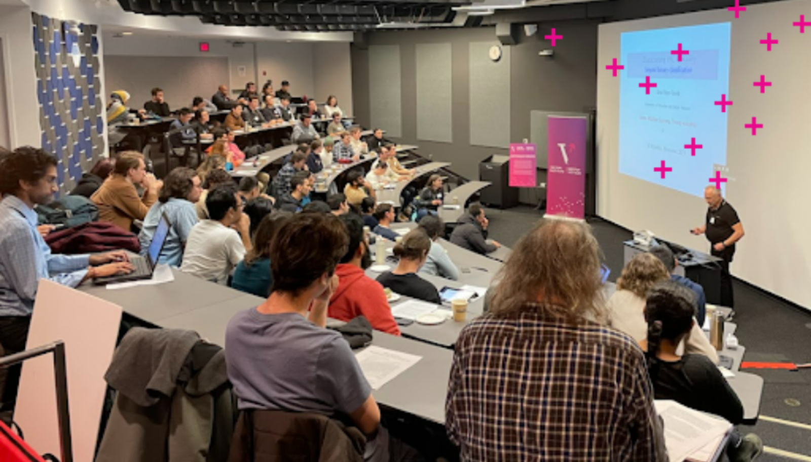
As senior editor for Medical Image Analysis and fellow of the Medical Image Computing and Computer Assisted Intervention (MICCAI) Society, Martel has observed a significant shift in medical imaging AI’s relationship to the broader computer science community. A decade ago, medical imaging researchers typically learned about new machine learning advances at conferences like ICCV, ICML, and NeurIPS, then adapted those methods to medical problems. The innovations flowed primarily from computer science into medicine.
That dynamic has changed. Increasingly, novel developments originate within medical imaging research itself. The U-Net architecture, now ubiquitous for image segmentation across many domains, was originally developed for and presented at MICCAI, the main medical imaging conference. Medical imaging researchers are now creating foundational methods, not just applying them. The field has matured from following methodological advances to contributing new approaches that influence the broader AI community.
This shift matters for institutions like Vector. Supporting domain-specific AI research in areas like medical imaging isn’t just about applying general techniques to new problems, it’s about recognizing that deep engagement with domain challenges often produces genuinely novel methodological innovations that benefit the entire field.
That editorial role also provides Martel with perspective on emerging challenges as AI becomes more prevalent. The volume of paper submissions has grown dramatically, and distinguishing truly innovative work from incremental modifications has become increasingly difficult. For a researcher focused on clinical applications, an additional filter is essential: does this work actually provide value in medical settings, or is it methodological innovation without practical applicability?
“I’m very much on the applied side. So you kind of have to ask yourself, well, this is great from a theory point of view, but is it ever going to be any use and is it appropriate? And also, have they tested it properly in a clinical domain?”
How Vector enables medical AI research
For researchers working at the boundary of computer science and medicine, institutions like Vector provide crucial infrastructure. Medical AI research requires genuine expertise in both domains – understanding both the technical capabilities of modern machine learning and the specific requirements and constraints of clinical applications.
Vector facilitates this bridging in several ways. The institute brings together researchers from computer science and various application domains, creating natural opportunities for collaboration and knowledge exchange. For medical imaging researchers, Vector provides access to AI engineering expertise that can help implement and scale methods, along with computational resources that make training large models feasible.The progression from Faculty Affiliate to Faculty Member provides increasingly deep integration into this ecosystem, with expanded resources and stronger collaborative connections that enable more ambitious research programs. For researchers considering whether Vector affiliation would benefit their work, the question is whether access to this interdisciplinary community, combined with computational and engineering support, would accelerate their research trajectory. For those genuinely pursuing work at disciplinary boundaries, the answer is often yes.